Mycosis fungoides symptoms – what to watch for
When dealing with Mycosis fungoides, a rare form of cutaneous T‑cell lymphoma that begins on the skin as flat patches and can evolve into raised plaques or tumors. Also known as cutaneous T‑cell lymphoma, it often masquerades as common skin problems, making early recognition crucial.
Key symptom patterns
The first clue usually appears as skin lesions, soft, reddish‑brown patches that may be scaly or slightly raised. These patches tend to linger for months, sometimes years, without healing. Many patients report a persistent itch that worsens at night, so itching, often described as a dry or burning sensation, becomes a hallmark symptom. The lesions often favor the trunk, buttocks, or inner thighs, but they can show up anywhere on the body.
As the disease moves beyond the patch stage, the skin changes take on a new look. Raised, thicker plaques replace the flat patches, and the color may darken to a deep bronze or violet hue. Some plaques develop a rough texture that feels like sandpaper. In later stages, nodules or tumors can form, sometimes ulcerating or bleeding. Because these transformations can look like eczema, psoriasis, or even a fungal infection, confusion is common. That's why understanding how mycosis fungoides symptoms differ from ordinary rashes or a fungal skin infection is essential for getting the right care.
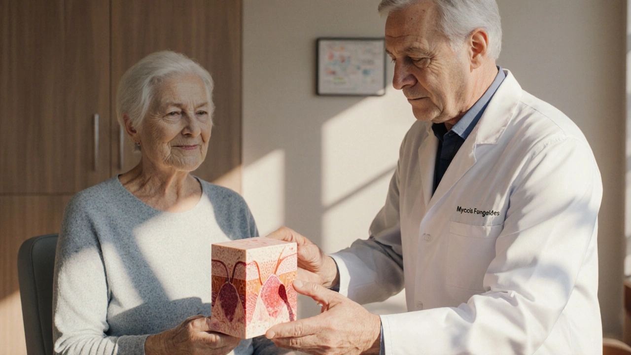
Getting a correct diagnosis hinges on a skin biopsy. During the procedure, a dermatologist removes a small sample of the suspicious area and sends it to a lab for histopathologic analysis. The pathologist looks for atypical T‑cells in the epidermis and dermis—a defining feature of Mycosis fungoides. Immunohistochemistry helps confirm the presence of CD4‑positive T‑cells and rule out other skin conditions. In short, skin biopsy, the gold‑standard test for confirming Mycosis fungoides, provides the definitive answer when visual clues alone aren’t enough.
Once the biopsy confirms the disease, doctors stage it based on skin involvement, lymph node status, blood involvement, and any visceral spread. Early‑stage disease (IA‑IIA) usually stays limited to the skin, while advanced stages (IIB‑IV) may involve the blood, lymph nodes, or internal organs. An advanced form called Sezary syndrome features circulating malignant T‑cells and generalized erythroderma. Recognizing the stage helps tailor treatment and predict prognosis. For most people, early detection means a broader range of skin‑directed therapies and a better quality of life.
Treatment choices depend on the stage and how aggressive the lesions appear. For patch‑stage disease, topical steroids, nitrogen mustard, or retinoids often provide relief. Phototherapy—especially narrow‑band UVB or PUVA—targets the abnormal T‑cells with light, slowing their growth. When plaques or tumors emerge, radiation therapy or systemic options like interferon‑alpha, histone deacetylase inhibitors, or newer targeted agents may be needed. The goal is always to control skin symptoms, reduce itching, and prevent progression to more serious forms.
The symptom story of Mycosis fungoides is a puzzle of lingering patches, stubborn itching, and evolving skin changes. By learning to spot the early clues, requesting a skin biopsy when doubts arise, and understanding how staging drives treatment, you empower yourself or a loved one to stay ahead of the disease. Below you’ll find articles that dive deeper into each of these areas—covering everything from differentiating fungal‑like rashes to the latest phototherapy protocols—so you can turn knowledge into action.
Mycosis Fungoides Myths: Facts, Truths & Real Treatment Insights
Clearly separates myths from facts about Mycosis Fungoides, explains staging, diagnosis, modern treatments, and offers practical advice for patients.